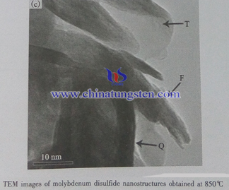
Molybdenum Disulfide TEM

Introduction
Transmission electron microscopy (TEM) is a microscopy technique in which a beam of electrons is transmitted through an ultra-thin specimen, interacting with the specimen as it passes through it. An image is formed from the interaction of the electrons transmitted through the specimen and the image is magnified and focused onto an imaging device, such as a fluorescent screen, on a layer of photographic film, or to be detected by a sensor such as a CCD camera.
The word “transmission” means “to pass through”. Essentially, the way the transmission electronmicroscope creates a conventional image (usually termed a bright field image) of a sample can be compared to shadow puppetry. Imagine a torch beam shone through a lattice on a window. The light passes through the transparent parts of the window, but is stopped by the lattice bars.The TEM uses a beam of highly energetic electrons instead of light from a torch. On the way through the sample some parts of the material stop or deflect electrons more than other parts. The electrons are collected from below the sample onto a phosphorescent screen or through a camera. In the regions where electrons do not pass through the sample the image is dark. Where electrons are unscattered, the image is brighter, and there are a range of greys in between depending on the way the electrons interact with and are scattered by the sample.
The left is the TEM image of molybdenum disulfide microparticle and the right one is the TEMimage of MoS2 nanostructure obtained at 850℃.

